Description
INTRODUCTION: Human respiratory syncytial virus (RSV) is an enveloped, nonsegmented, negative-sense, single-stranded RNA virus belonging to the Pneumovirus genus of the subfamily Pneumovirinae, the family Paramyxoviridae. RSV is the most common virus responsible for acute and severe lower airway disease in infants and young children worldwide. Despite the enormous burden of RSV disease, there is no efficacious vaccine or antiviral drug therapy yet available. The RSV genome (15.2 kb) contains 10 mRNAs encoding 11 proteins. The nucleocapsid (N) protein binds the negative-strand RNA genome and associates with the phosphoprotein (P), the large (L) polymerase protein, and the M2-1 protein to form the nucleocapsid. The matrix (M) protein is present between the nucleocapsid and the outer envelope and plays a structural role in virion assembly and budding. There are three envelope glycoproteins: the attachment glycoprotein (G), the fusion (F) protein, and the small hydrophobic (SH) protein. The genome also encodes two nonstructural proteins (NS1, NS2) which suppress the interferon response and M2-2 protein (the second product of the M2 gene) which governs the transition from transcription to replication of genomic RNA.
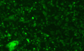
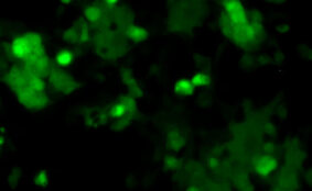
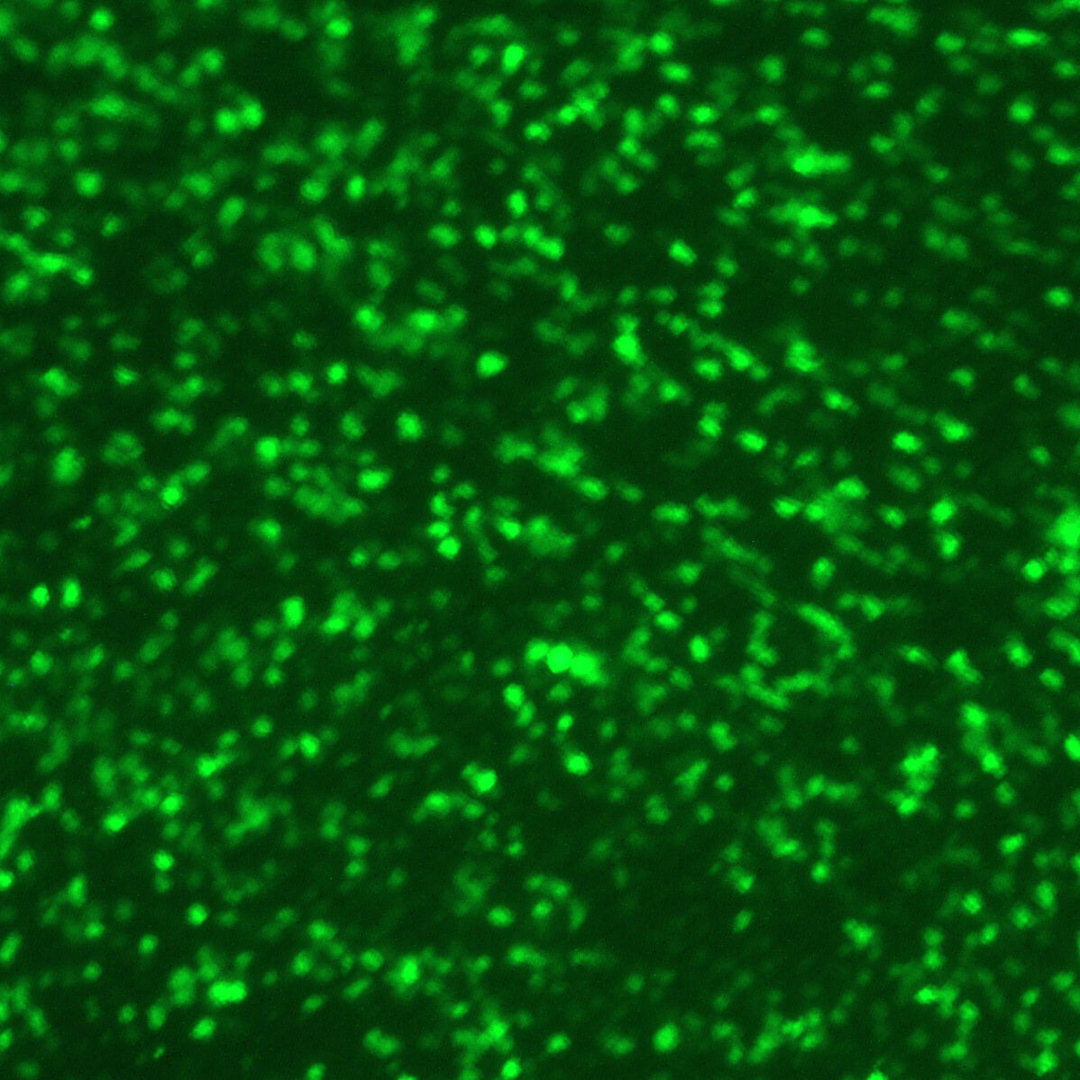
DESCRIPTION: Recombinant RSV with both green fluorescent protein (GFP) and luciferase (Luc) was constructed to contain the GFP (Green Lantern Protein, Life Technologies) and Renilla luciferase (Promega) as its first and second genes in front of NS1 gene (RSV-GFP1-Luc2). Both GFP and Luc genes were amplified by PCR with primers that added RSV transcription signals before being inserted into the full-length anti-genomic plasmid. RSV-GFP1-Luc2 was rescued by cotransfecting HEp-2 cells with the antigenomic plasmid and N, P, M2-1, and L support plasmids and infecting them with a modified vaccinia virus, MVA-T7, expressing T7 RNA polymerase. Of note, the recovered virus contained a spontaneous mutation in its G gene (deletion of an A in codon 66), resulting in a frame shift that truncated its ectodomain, while the N-terminal cytoplasmic and transmembrane domains of G were intact. This virus replicated to a lower titer as compared to its parental virus.
| Parental Strain: | A2 strain |
| Construction: | GFP and Luc genes were inserted in front of NS1 gene (as the 1st & 2nd gene). |
| Passage History: | The isolate was plaque purified and propagated in HEp-2 cells. |
| Infectivity: | Titer > 5.7 log10 TCID50 per mL. Infectious in humans. |
| Volume/Storage: | 2 x 1.2 mL per cryovial. Store at –80ºC. |
| Quality Testing: | No bacteria, fungus, or mycoplasma detected. Endotoxin <10 EU/mL. |
| Availability: | Bulk quantity and custom orders are available. Contact info@viratree.com. |
Product sheet: Product_R141
Certificate of Analysis: CoA_Lot1761
Material Safety Data Sheet (MSDS): MSDS_RSV