Description
INTRODUCTION: Sendai virus (SeV), also known as murine parainfluenza virus type 1 or hemagglutinating virus of Japan (HVJ), is a nonsegmented negative sense single-stranded RNA virus of the Respirovirus genus of the family Paramyxoviridae. SeV is one of the most important respiratory pathogens of rats and mice. SeV is responsible for a highly transmissible respiratory tract infection in mice, hamsters, guinea pigs, rats, and occasionally pigs, with infection passing through both air and direct contact routes. Clinical symptoms seen in mice are signs of pneumonia, such as dyspnea, chattering teeth, and death in young mice. Multiple strains of SeV have been described. The SeV Z strain has a 15,384-nucleotide RNA genome, consisting of six genes. Each gene has concise transcription initiation and termination signals and is transcribed to mRNA encoding a single polypeptide (except for the P gene encoding P, C and V proteins). SeV particles are spherical in shape with an average diameter of 260 nm. A SeV virion consists of the nucleocapsid (genomic RNA complexed with NP, P and L proteins), an envelope (a lipid bilayer with F and HN proteins) and a matrix (M protein) connecting the nucleocapsid and envelope. Since its discovery, SeV has been widely used as a research tool in cell biology and immunology. Because of its low pathogenicity in humans and a wide host range, SeV has been increasingly used as a cytoplasmic gene delivery/expression vector in applications such as gene therapy and regenerative medicine.
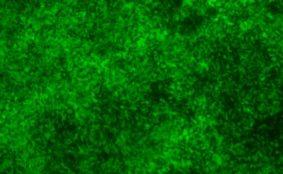
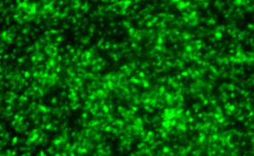
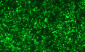
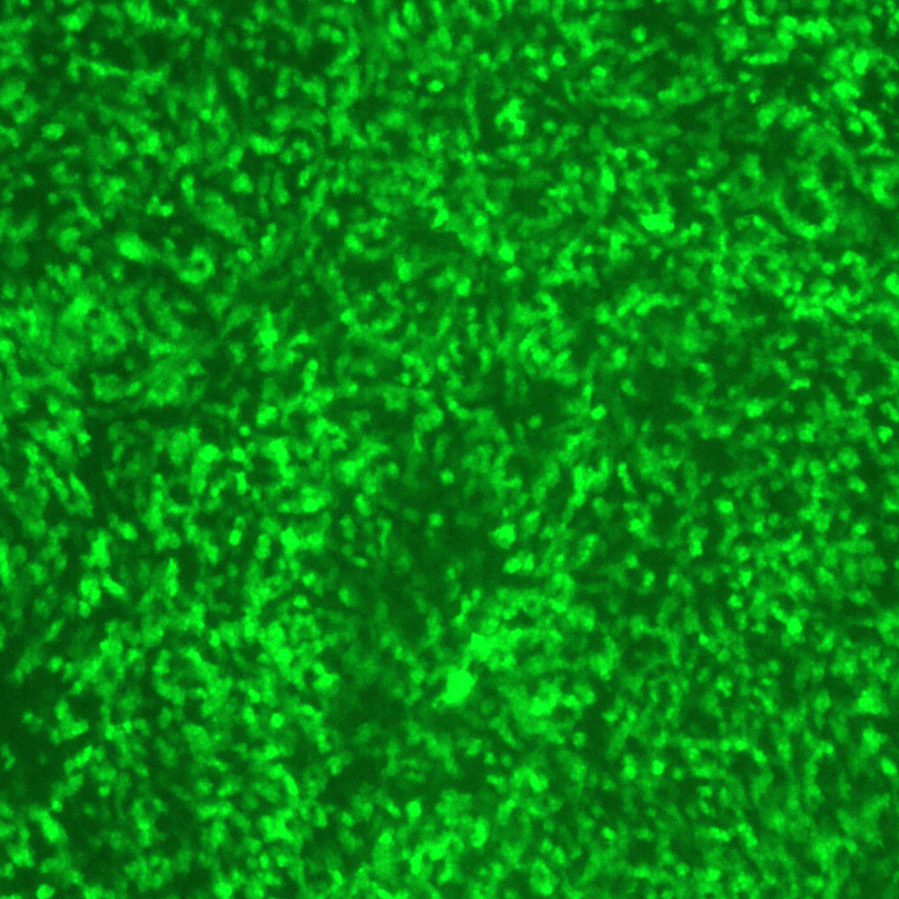
DESCRIPTION: The full-length clone of wild-type SeV of the Z strain, pFL5, was constructed to contain a unique MluI restriction site and an insert of 30 nucleotides containing extra transcriptional signals at position 4832 in the 3’ untranslated region of the F gene. The GFP gene sequence was introduced in the MluI site between the M and F genes to generate the pFL5-GFP clone. To rescue the recombinant SeV expressing GFP (SeV-GFP4), BSR-T7 cells were transfected with the full-length pFL5-GFP plasmid and pTM1-based support plasmids (harboring an encephalomyocarditis virus internal ribosomal entry site) expressing the N, P/C, and L genes. Twenty-four hours posttransfection, the cells were collected and injected into 9-day-old embryonated chicken eggs. After three days incubation at 33°C, the allantoic fluids were collected. SeV-GFP4 was further amplified through egg-to-egg passages. Infectious viral stocks were finally prepared in LLC-MK2 cells.
| Parental Strain: | Wild-type Z strain |
| Construction: | GFP gene was inserted between M and F genes as the 4th gene. |
| Passage History: | The isolate was finally propagated in LLC-MK2 cells. |
| Infectivity: | Titer > 7.0 log 10 TCID50 per mL. |
| Volume/Storage: | 2 x 1.2 mL per cryovial. Store at –80ºC. |
| Quality Testing: | No bacteria, fungus, or mycoplasma detected. Endotoxin <10 EU/mL. |
| Availability: | Bulk quantity and custom orders are available. Contact info@viratree.com. |
Product Sheet: Product_S124
Certificate of Analysis: CoA_Lot1548&1549
Material Safety Data Sheet (MSDS): MSDS_PIV